Travel inside the eyes — our window to the world — and learn how they allow us to see objects both far and near.
How The Eye Works
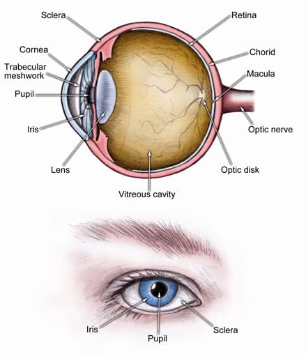
In order to see, there must be light. Light reflects on an object and — if one is looking at the object — enters the eye.
The first thing light touches when entering the eye is a thin veil of tears that coats the front of the eye. Behind this lubricating moisture is the front of the eye, called the cornea. This clear covering helps to focus the light.
On the other side of the cornea is more moisture. This clear, watery fluid is the aqueous humor. It circulates throughout the front part of the eye and keeps a constant pressure within the eye.
How The Iris Is Effected
After light passes through the aqueous humor, it passes through the iris. This is the colored part of the eye. Depending on how much light there is, the iris may contract or dilate, limiting or increasing the amount of light that gets deeper into the eye. After light flows through the iris it enters the pupil — the black dot in the middle of the eye. The light then goes through the lens. Just like the lens of a camera, the lens of the eye focuses the light. The lens changes shape to focus on light reflecting from near or distant objects.
This focused light now beams through the center of the eye. Again the light is bathed in moisture, this time in a clear jelly known as the vitreous. Surrounding the vitreous is the tough, fibrous, white part of the eye known as the sclera. It protects the delicate structures inside the eye.
What Happens When The Light Reaches The Retina
At last the light reaches its final destination: the retina located at the back of the eye. In a way, the retina is like a movie screen. The focused light is projected onto its flat, smooth surface. However, unlike a movie screen, the retina has many working parts:
- Blood vessels. Behind the retina is a layer of blood vessels called the choroids that bring nutrients to the retina.
- The macula. This is the bull’s-eye at the center of the retina. The dead center of this bull’s eye is called the fovea. Because it’s at the focal point of the eye, it has more specialized, light sensitive nerve endings, called photoreceptors, than any other part of the retina.
- Photoreceptors. There are two kinds of photoreceptors: rods and cones. These specialized nerve endings convert the light into electro-chemical signals.
- Retinal pigment epithelium. Beneath the photoreceptors is a layer of dark tissue known as the retinal pigment epithelium, or RPE. These important cells absorb excess light so that the photoreceptors can give a clearer signal. They also move nutrients to (and waste from) the photoreceptors to the choroid. Bruch’s membrane separates the choroid from the RPE.
Signals sent from the photoreceptors travel along nerve fibers to a nerve bundle at the back of the eye, called the optic nerve. It carries all the information collected from the eye to the brain.
Now light has reflected from an object, entered the eye, been focused, and converted into electro-chemical signals. But seeing hasn’t yet happened. That’s because the eye is only part of the story. Now the brain must receive — and interpret — the eye’s signals. Once this is done, vision occurs.